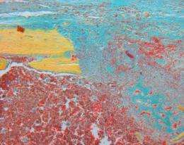
Technique triggers rapid regrowth in damaged bone (Update)

(PhysOrg.com) -- Imagine being able to re-grow a broken bone three times more quickly than normal. (Harry Potter fans? Think Skele-gro.) That’s just what researchers at the Stanford University School of Medicine have done in laboratory mice.
They stimulated the rapid bone growth by injecting a protein called Wnt known to be involved in the growth of many types of tissues in animals like salamanders, zebrafish and mice. The feat marks the first time that researchers have managed to package the Wnt protein in a form that could be used in humans, and opens the door to additional experiments to heal skin, muscle, brain and other tissue injuries.
“We believe our strategy has the therapeutic potential to accelerate and improve tissue healing in a variety of contexts,” said Jill Helms, DDS, PhD, professor of surgery. “There is an enormous amount of literature about the role of Wnt in tissue growth, but until now we’ve not been able to directly test whether Wnt proteins could aid regeneration in mammals.”
It may also eventually provide a much needed alternative to currently available drugs based on bone morphogenetic proteins, or BMPs. BMPs have been approved for use in humans to speed bone growth in spinal fusions and long bone fractures, but they’ve become increasingly associated with a number of adverse side effects.
Helms is the senior author of the work, published April 28 in the online journal Science Translational Medicine. The work represents an ongoing collaboration with Stanford developmental biology professor Roel Nusse, PhD. Nusse was the first to identify the Wnt protein in 1982, and in 2002 his lab was the first to purify the protein in an active form. These accomplishments set the stage for this study.
Researchers have known for years that Wnt is a key player in tissue regeneration in many organisms, and speculation about its possible therapeutic uses in humans has abounded. But the protein proved to be a cantankerous research subject. It was exceedingly difficult to purify in an active form and, once isolated, was found to be both insoluble and hydrophobic (meaning it’s difficult to dissolve in nearly any liquid). Basically, it doesn’t play well with others. As a result, researchers have been stymied as to how to test its effects in living animals.
Helms, Nusse and their collaborators — including medical student Steve Minear, orthopedic surgery resident Philipp Leucht, MD, and postdoctoral scholar Jie Jiang, PhD — overcame this problem by using tiny, water-friendly molecular bubbles called liposomes. Liposomes are made up of the same molecules as a cell membrane, and they are sometimes used to surround and deliver chemotherapy drugs to tumors. In this case, however, the researchers planted laboratory-made Wnt on the bubble’s outer surface like a flag. The Wnt-adorned liposomes can then be suspended in liquid for delivery into the body.
“We took advantage of the way the protein is made in nature to present it in what turned out to be a highly active form,” said Helms. “This allowed us to start testing its activity in animals.”
To do so, Helms and her colleagues drilled 1-millimeter holes into the leg bones of the mice with a high-speed dental drill. (The animals were anesthetized during the procedure and given painkillers while healing.) They then injected the Wnt-covered liposomes at the site of the damage and compared the rate and pattern of bone formation with that of control animals and with animals injected with Wnt without a liposomal escort.
Within three days, the animals who had received Wnt had 3.5 times more new bone than the other two groups. And after 28 days the newly formed bone had completely healed, while the control animals were still in the process of repairing the injury. Further investigation showed that Wnt works by increasing the proliferation of bone progenitor cells, which then became new bone.
In many ways, the Wnt-activated healing mimics what happens naturally. Wnt-responsive cells are found on the inner surface of the bone, and they participate in normal bone maintenance and in bone growth in response to injury. In fact, Helms and Nusse found that laboratory mice that had been genetically modified to have a more prolonged Wnt signal also healed more quickly than did control mice. They also confirmed that, as in other animals, the Wnt pathway is activated in response to injury in mice and is required for normal healing.
The researchers plan to continue their studies of the Wnt pathway in other tissues. Intriguingly, flatworms and zebrafish rely on the Wnt pathway to regenerate lost tissue without scarring. Capturing that ability in humans — perhaps by applying extra Wnt at a particular time and place in the body — could improve our ability to heal.
“After stroke and heart attack,” said Helms, “we heal the injuries slowly and imperfectly, and the resulting scar tissue lacks functionality. Using Wnt may one day allow us to regenerate tissue without scarring.” These studies are currently ongoing at the medical school in the laboratories of Theo Palmer, PhD, associate professor of neurosurgery, and Joseph Wu, MD, PhD, assistant professor of medicine and of radiology.
More information: "Wnt Proteins Promote Bone Regeneration," by S. Minear; Y.A. Zeng; C. Fuerer; R. Nusse at Howard Hughes Medical Institute in Stanford, CA; S. Minear; P. Leucht; J. Jiang; B. Liu; Y.A. Zeng; C. Fuerer; R. Nusse; J.A. Helms at Stanford University School of Medicine in Stanford, CA.


